
Ibidi – Labware – µ-Slide Membrane ibiPore Flow
Product Outline:
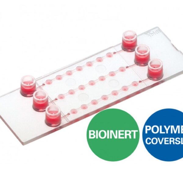
- A µ-Slide with a porous glass membrane for transmigration and transport studies under both static and flow conditions
- Brilliant optical quality throug...
Product Outline:
- A µ-Slide with a porous glass membrane for transmigration and transport studies under both static and flow conditions
- Brilliant optical quality through the thin, porous glass membrane
- Full access to the apical and basal sides of adherent cells in numerous applications
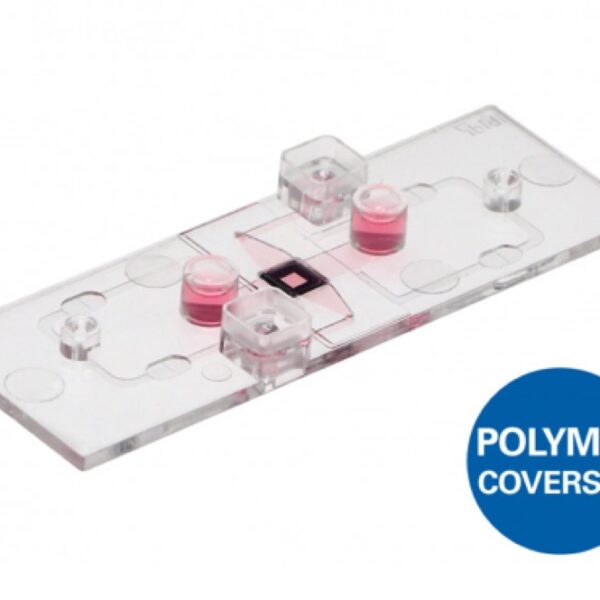
- Different pore sizes available for various cell types
- Suitable for the establishment of lung models with air-liquid interface (ALI)
Applications
- Trans-endothelial migration under flow conditions
- Co-cultivation of cell layers and transport in 2D or in a 3D gel matrix
- Apical-basal cell polarity assays
- Skin and lung models with liquid-air interface
- Cell barrier model assays with apical-basal gradients
- Cell migration assays based on filters and porous membranes