Showing 197–200 of 405 results

  • Ibidi - Labware - µ-Slide Angiogenesis

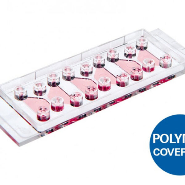
    Ibidi – Labware – µ-Slide Angiogenesis

    Product outline:

    • A µ-Slide used to investigate angiogenesis in tube formation assays. Also ideal for 3D cell culture and immunofluorescence staining.
    • Complete solu...

    Product outline:

    • A µ-Slide used to investigate angiogenesis in tube formation assays. Also ideal for 3D cell culture and immunofluorescence staining.
    • Complete solution for tube formation experiments, requiring only a few steps from sample preparation to image analysis
    • Brilliant visualization without meniscus formation, and with all cells in one focal plane
    • Can be used with a broad range of gels (e.g., Matrigel®, collagen, and agarose)
    • Cost-effective experiments, requiring only 10 µl of gel per well

    Applications

    • Tube formation assays
    • 3D cell culture
    • Immunofluorescence staining
    • Sprouting assays
    • Live cell imaging of attached cells
  • Ibidi - Labware - µ-Slide Chemotaxis

    Ibidi – Labware – µ-Slide Chemotaxis

    Product outline:

    • Investigate chemotaxis of fast or slow migrating adherent cells and non-adherent cells in 2D or 3D gel matrices
    • Real-time chemotaxis measurement in r...

    Product outline:

    • Investigate chemotaxis of fast or slow migrating adherent cells and non-adherent cells in 2D or 3D gel matrices
    • Real-time chemotaxis measurement in real-time in a 2D or 3D environment
    • Stable chemotactic gradients for long-term experiments
    • Reproducible results with reliable and user-independent data

    Applications

    • 2D and 3D chemotaxis assays with fast or slow migrating cells
    • Live cell imaging using inverted microscopy
    • Chemotaxis of neutrophils, lymphocytes, and monocytes
    • 3D chemotaxis of leukocytes or cancer cells in an ECM-like matrix
    • Invasion assays of tumour cells in Matrigel™
    • Chemotaxis measurements of adherent and non-adherent cells
  • Ibidi - Labware - µ-Slide I Luer

    Ibidi – Labware – µ-Slide I Luer

    Product outline:

    • Channel slides with different heights, volumes, and coatings especially suited for flow applications
    • Large area of uniform shear stress
    • Easy ...

    Product outline:

    • Channel slides with different heights, volumes, and coatings especially suited for flow applications
    • Large area of uniform shear stress
    • Easy connection using Luer adapters
    • Homogeneous cell distribution with defined optical pathway

    Applications

    • Adherent cells under flow conditions
    • Cell culture (static or stop-flow)
    • Simulation of blood vessels with endothelial cells
    • High-resolution microscopy of living and fixed cells
  • Ibidi - Labware - µ-Slide I Luer 3D

    Ibidi – Labware – µ-Slide I Luer 3D

    A µ-Slide With One Channel and Three Wells for Culturing Cells on a 3D Gel Matrix With Defined Flow

    Product outline:

    • Create a perfusable cell monolayer ...

    A µ-Slide With One Channel and Three Wells for Culturing Cells on a 3D Gel Matrix With Defined Flow

    Product outline:

    • Create a perfusable cell monolayer on a gel matrix for various flow and shear stress applications
    • Establish and image endothelial barriers under in vivo-like conditions—no artificial filters or membranes
    • Easy handling: samples are placed into the open wells; perfusion is applied after closing the wells

    Applications

    • Culture of adherent cells on a 3D gel matrix under flow conditions
    • Transendothelial migration studies under flow conditions
    • Establishment and microscopic read-out of endothelial barrier assays (without any artificial membrane)
    • Simulation of blood vessels with endothelial cell culture on a soft substrate
    • Long-term or short-term cell culture perfusion experiments with defined shear stress and optimal nutrient supply into 3D structures
    • Apical-basal cell polarity assays
    • Cell barrier model assays with apical-basal gradients
    • Cell boundary assays without filters or porous membranes
    • Rolling and adhesion assays using leukocytes
    • Inflammation studies
    • Transport studies