Showing 13–16 of 16 results

  • Ibidi - Labware - µ-Slide I Luer

    Ibidi – Labware – µ-Slide I Luer

    Product outline:

    • Channel slides with different heights, volumes, and coatings especially suited for flow applications
    • Large area of uniform shear stress
    • Easy connection using Luer adapters
    • Homogeneous cell distribution with defined optical pathway

    Applications

    • Adherent cells under flow conditions
    • Cell culture (static or stop-flow)
    • Simulation of blood vessels with endothelial cells
    • High-resolution microscopy of living and fixed cells
  • Ibidi - Labware - µ-Slide I Luer 3D

    Ibidi – Labware – µ-Slide I Luer 3D

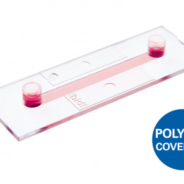
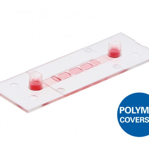
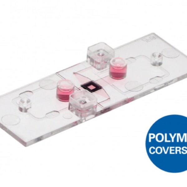
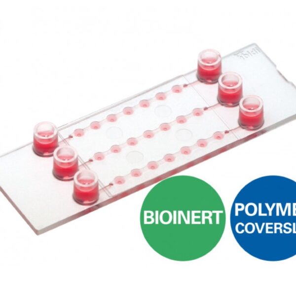
    A µ-Slide With One Channel and Three Wells for Culturing Cells on a 3D Gel Matrix With Defined Flow

    Product outline:

    • Create a perfusable cell monolayer on a gel matrix for various flow and shear stress applications
    • Establish and image endothelial barriers under in vivo-like conditions—no artificial filters or membranes
    • Easy handling: samples are placed into the open wells; perfusion is applied after closing the wells

    Applications

    • Culture of adherent cells on a 3D gel matrix under flow conditions
    • Transendothelial migration studies under flow conditions
    • Establishment and microscopic read-out of endothelial barrier assays (without any artificial membrane)
    • Simulation of blood vessels with endothelial cell culture on a soft substrate
    • Long-term or short-term cell culture perfusion experiments with defined shear stress and optimal nutrient supply into 3D structures
    • Apical-basal cell polarity assays
    • Cell barrier model assays with apical-basal gradients
    • Cell boundary assays without filters or porous membranes
    • Rolling and adhesion assays using leukocytes
    • Inflammation studies
    • Transport studies
  • Ibidi - Labware - µ-Slide Membrane ibiPore Flow

    Ibidi – Labware – µ-Slide Membrane ibiPore Flow

    Product Outline:

    • A µ-Slide with a porous glass membrane for transmigration and transport studies under both static and flow conditions
    • Brilliant optical quality through the thin, porous glass membrane
    • Full access to the apical and basal sides of adherent cells in numerous applications
    • Different pore sizes available for various cell types
    • Suitable for the establishment of lung models with air-liquid interface (ALI)

    Applications

    • Trans-endothelial migration under flow conditions
    • Co-cultivation of cell layers and transport in 2D or in a 3D gel matrix
    • Apical-basal cell polarity assays
    • Skin and lung models with liquid-air interface
    • Cell barrier model assays with apical-basal gradients
    • Cell migration assays based on filters and porous membranes
  • Ibidi - Labware - µ-Slide Spheroid Perfusion

    Ibidi – Labware – µ-Slide Spheroid Perfusion

    A Perfusable Channel µ-Slide With 3 x 7 Wells for Long-Term Spheroid Cultivation and High-End Microscopy

    Product outline:

    • Ideal for the long-term cultivation and perfusion of 3D spheroids or organoids on the Bioinert surface
    • Flexible preparation for perfusion: spheroids can be transferred or created directly in the wells
    • Imaging chamber with excellent optical quality for high-resolution microscopy

    Applications

    • Cultivation and high-resolution microscopy of three-dimensional cell aggregates (e.g., spheroids and organoids)
    • For use with the ibidi Pump System or any other pump device with Luer connectors
    • Perfusion of spheroids for fresh medium supply during long-term cultivation and access to spheroid growth kinetics
    • Spheroid generation directly in the wells
    • Spheroid retrieval for downstream processing (e.g., immunofluorescence, histology, and biochemical assays)
    • Perfusion of cell monolayers (adherent cells), suspension cells, or co-cultures
    • Organ-on-a-chip setups with up- and downstream metabolization
    • Live cell imaging and microscopy of 3D cell aggregates
    • Immunofluorescence staining and high-resolution fluorescence microscopy of living and fixed cells and cell aggregates